Accessory (OPA)

Optics Plate Accessory (OPA)
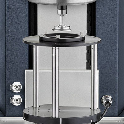
The OPA is an open optical system that permits basic visualization of sample structure during rheological experiments, revealing important insights about material behavior under flow. An open platform with a borosilicate glass plate provides a transparent optical path through which the sample can be viewed directly. This enhances the understanding of a range of materials, especially suspensions and emulsions. The accessory is easy to use and install, accommodates diverse optical systems, and offers accurate temperature control over a wide range for flow visualization and microscopy.
Features and Benefits
- Smart Swap™ technology for quick installation
- Simultaneous rheological measurements and direct visualization
- Visual access to any position within the measurement area, e.g. center, edge, or mid-radius
- Upper Peltier Plate (UPP) with patented Active Temperature Control for precise temperature measurement
Technology
The OPA mounts to the DHR Smart SwapTM base and may be coupled with the Upper Peltier Plate with Active Temperature Control for accurate, direct sample temperature measurement and control from -20 °C to 100 °C. The OPA can be used with cone or parallel plate geometries up to 60 mm in diameter.
The OPA is available in any of the following configurations:
- Open Plate: An open system that facilitates customization including a set of 8 M2 tapped holes for the easy adaptation of any optical system
- OPA with Modular Microscope Accessory (MMA): A static optical stage for microscopy.
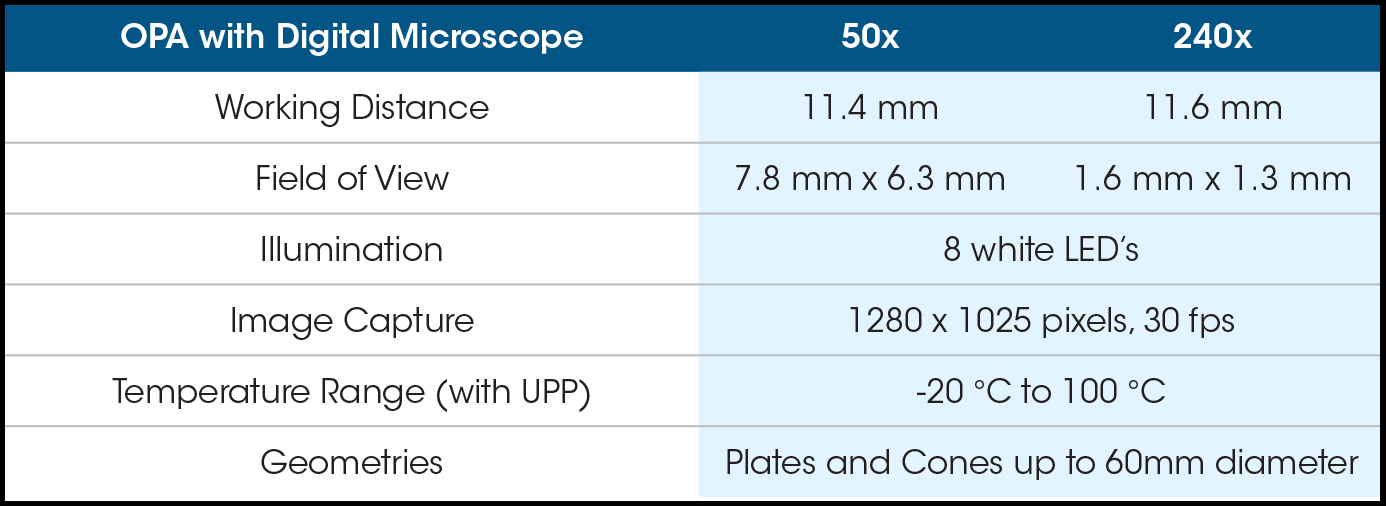
- OPA with Digital Microscope: A high resolution digital camera permits the capture of still images or video. The camera is mounted on a y-z positioning stage to adjust focus and the field of view. Sample illumination is provided by the microscope’s 8 white LEDs.
Microscopy
OPA with Digital Microscope

OPA with Digital Microscope
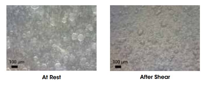
The images below show the structure of a PDMS-PIB emulsion at rest and after shear flow. At rest, the emulsion structure consists of spherical droplets with a polydisperse size distribution. After shearing at 10 s-1 for 10 minutes, there is a decrease in the number of larger droplets and a shift towards more uniform droplet sizes.

- Description
-
Optics Plate Accessory (OPA)
The OPA is an open optical system that permits basic visualization of sample structure during rheological experiments, revealing important insights about material behavior under flow. An open platform with a borosilicate glass plate provides a transparent optical path through which the sample can be viewed directly. This enhances the understanding of a range of materials, especially suspensions and emulsions. The accessory is easy to use and install, accommodates diverse optical systems, and offers accurate temperature control over a wide range for flow visualization and microscopy.
- Features
-
Features and Benefits
- Smart Swap™ technology for quick installation
- Simultaneous rheological measurements and direct visualization
- Visual access to any position within the measurement area, e.g. center, edge, or mid-radius
- Upper Peltier Plate (UPP) with patented Active Temperature Control for precise temperature measurement
- Technology
-
Technology
The OPA mounts to the DHR Smart SwapTM base and may be coupled with the Upper Peltier Plate with Active Temperature Control for accurate, direct sample temperature measurement and control from -20 °C to 100 °C. The OPA can be used with cone or parallel plate geometries up to 60 mm in diameter.
The OPA is available in any of the following configurations:
- Open Plate: An open system that facilitates customization including a set of 8 M2 tapped holes for the easy adaptation of any optical system
- OPA with Modular Microscope Accessory (MMA): A static optical stage for microscopy.
- OPA with Digital Microscope: A high resolution digital camera permits the capture of still images or video. The camera is mounted on a y-z positioning stage to adjust focus and the field of view. Sample illumination is provided by the microscope’s 8 white LEDs.
- Applications
-
Microscopy
OPA with Digital Microscope

OPA with Digital Microscope
The images below show the structure of a PDMS-PIB emulsion at rest and after shear flow. At rest, the emulsion structure consists of spherical droplets with a polydisperse size distribution. After shearing at 10 s-1 for 10 minutes, there is a decrease in the number of larger droplets and a shift towards more uniform droplet sizes.


